Dr. MONTANA SUPAWONGWATTANA
Ophthalmology



A gel-like substance inside the eye, located behind the lens. It plays a role in maintaining the shape of the eyeball. Its key components include water, fibrous proteins like collagen, hyaluronic acid, and various minerals.
The vitreous is clear and free of debris, adhering closely to the back of the lens and the front of the retina. However, as people reach middle age or encounter other contributing factors, the vitreous begins to degenerate. The fibrous proteins can break into tiny strands or clump together, reducing the vitreous's transparency.
This leads to symptoms such as seeing dark shadows, which may appear as small dots, lines, watermarks, cobweb-like patterns, or rings that float with eye movement. These are more noticeable in certain lighting conditions, such as looking at a bright sky, a white wall, or reading in a well-lit environment.

Dark spots or cobweb-like strands that float within your vision. These may become more noticeable when you move your eyes or look at a white surface or the sky.
Caused by the vitreous pulling on or detaching from the retina, stimulating the retina, and creating a sensation of flashes of light. These flashes can resemble camera flashes or lightning streaks in the eye, even when the eyes are closed.
When the vitreous detaches or shrinks, it may pull tightly on blood vessels it adheres to, causing them to rupture. This leads to a sudden decline in vision, resembling dense fog or bright red vision due to blood.
Occurs when the vitreous is more firmly attached to certain areas of the retina. As the vitreous degenerates and contracts, it may tear the retina in these areas.
If a retinal tear worsens, the vitreous may pull the retina further, leading to detachment. This can cause symptoms like a dark curtain or shadow covering the field of vision. Immediate medical attention is required for diagnosis and treatment.
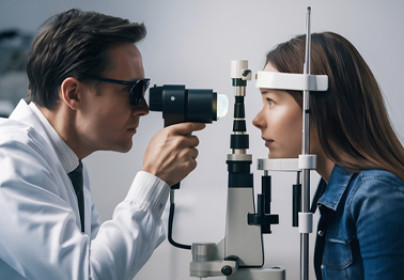
If any of the above symptoms occur, it is essential to consult an ophthalmologist for an eye examination. This includes dilation of the pupils using eye drops to allow a thorough assessment of the retina and vitreous. After dilation, vision may become blurry, and sensitivity to light increases, along with difficulty judging distances or focusing on close objects for about 4-6 hours.
Patients are advised not to drive themselves, bring a companion, and wear sunglasses to reduce light sensitivity.
Vitreous degeneration typically does not require treatment. While the symptoms can be bothersome, they usually do not cause severe harm. Over time, as the vitreous separation completes, debris within the vitreous may gradually diminish, and floaters or dark spots may fade. This process can take months to years, depending on the individual.
Be vigilant for new or worsening symptoms, such as bright red vision, frequent flashes of light, or shadows obscuring the field of vision. These signs may indicate complications involving the retina or blood vessels and require prompt treatment.
If a retinal tear or detachment occurs, treatments like laser therapy, cryopexy, or vitreoretinal surgery may be necessary. Follow-up appointments as recommended by the ophthalmologist are crucial after treatment.
Annual eye exams are recommended to detect and treat eye conditions early, thus preventing potential vision loss. As the risk of eye diseases increases with age, it's important to consult an ophthalmologist immediately if you notice any abnormalities.
For more information, please contact:
 Ophthalmology Department, 1st Floor, RAM 2 Hospital
Email: information@ram2-hosp.com
Line Official: @ram2
Tel: 02-032-3888
Ophthalmology

จักษุวิทยา (Ophthalmology)
จักษุวิทยา (Ophthalmology)